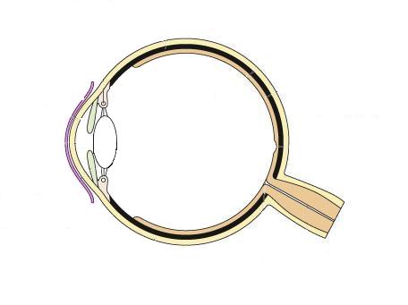
41 diagram of the human eye without labels
Eye Anatomy: 16 Parts of the Eye & Their Functions - Vision Center The lens of the eye (or crystalline lens) is the transparent lentil-shaped structure inside your eye. This is the natural lens. It is located behind the iris and to the front of the vitreous humor (vitreous body). The vitreous humor is a clear, colorless, gelatinous mass that fills the gap between the lens and the retina in the eye. label the eye anatomy diagram eye diagram label anatomy brain blank printable science clipart cliparts without structure experiments library activities rudyard class fair chart. LAS Assignment 7 - Eye Enucleation cmapsconverted.ihmc.us. eye cow anatomy human eyes dissection enucleation tapetum lucidum ihmc science rid. Human Eye Diagrams With The Unlabeled
File:Schematic diagram of the human eye en.svg - Wikimedia Diagram of the human eye in English. It shows the lower part of the right eye after a central and horizontal section. ... Full redraw: Group labels in accordance with the "Foundational Model Explorer." Added "Macula" and "Uvea" and removed "Zonular fibres." ... File:Diagram of human eye without labels.svg; File:Figure of diplopia perception ...

Diagram of the human eye without labels
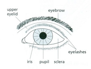
The Human Eye Worksheets | Human eye diagram, Parts of the ... - Pinterest Description Use these simple eye diagrams to help students learn about the human eye. Three differentiated worksheets are included: 1. Write the words using a word bank 2. Cut and paste the words 3. Write the words without a word bank Labels include: eyebrow, eyelid, eyelashes, pupil, iris, and sclera. UPDATE: I've updated this product to ... Eye Anatomy: Parts of the Eye and How We See Behind the anterior chamber is the eye's iris (the colored part of the eye) and the dark hole in the middle called the pupil. Muscles in the iris dilate (widen) or constrict (narrow) the pupil to control the amount of light reaching the back of the eye. Directly behind the pupil sits the lens. The lens focuses light toward the back of the eye. Eye Diagram Teaching Resources | Teachers Pay Teachers The Human Eye Overview Reading Comprehension and Diagram Worksheet. by. Teaching to the Middle. 4.7. (65) $1.50. Zip. This passage briefly describes the human eye (900-1000 Lexile). 14 questions (matching and multiple choice) assess students' understanding. Students label a diagram of 6 parts of the eye.
Diagram of the human eye without labels. Human Body Diagram - Bodytomy ☛ The human eye has the ability to differentiate between 400+ shades of gray, and what's more, it can identify approximately 10 million colors. ☛ Your ears never sleep. Sound is received even while you are asleep; it's the brain that does not process them. The Eyes (Human Anatomy): Diagram, Optic Nerve, Iris, Cornea ... - WebMD The front part (what you see in the mirror) includes: Iris: the colored part. Cornea: a clear dome over the iris. Pupil: the black circular opening in the iris that lets light in. Sclera: the ... Anatomy of the eye: Quizzes and diagrams | Kenhub Take a look at the diagram of the eyeball above. Here you can see all of the main structures in this area. Spend some time reviewing the name and location of each one, then try to label the eye yourself - without peeking! - using the eye diagram (blank) below. Unlabeled diagram of the eye Eye diagram by Firkin | Human eye diagram, Diagram of the eye, Eye ... Eye diagram by Firkin Find this Pin and more on free images to print by Monica Eggleton. Human Eye Diagram Diagram Of The Eye Eye Parts Parts Of The Eye Eye Anatomy Human Body Anatomy Eye Structure Eyes Clipart Free Human Body More information ... More information Eye diagram by Firkin
What Does the Eye Look Like? - Diagram of the Eye | Harvard Eye Associates Vitreous Gel: A thick, transparent liquid that fills the center of the eye. It is mostly water and gives the eye its form and shape. Our eyes are vital for seeing the world around us. Keep them healthy by maintaining regular vision exams. Contact Harvard Eye Associates at 949-951-2020 or harvardeye.com to schedule an appointment today. Structure and Functions of Human Eye with labelled Diagram - BYJUS The human eye is a roughly spherical organ, responsible for perceiving visual stimuli. It is enclosed within the eye sockets in the skull and is anchored down by muscles within the sockets. Anatomically, the eye comprises two components fused into one; hence, it does not possess a perfect spherical shape. Cross sectional diagram of human eye [1]. - ResearchGate A total of 100 photos were included in the analysis—50 sick and 50 normal eyes. Small lesions in diabetic retinopathy could be automatically diagnosed by the system with an accuracy of 98%. Human Ear Diagram - Bodytomy The Structure of Human Ear. Helix: It is the prominent outer rim of the external ear. Antihelix: It is the cartilage curve that is situated parallel to the helix. Crus of the Helix: It is the landmark of the outer ear, situated right above the pointy protrusion known as the tragus. Auditory Ossicles: The three small bones in the middle ear ...
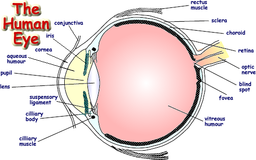
Eye Anatomy: A Closer Look At the Parts of the Eye - All About Vision In a number of ways, the human eye works much like a digital camera: Light is focused primarily by the cornea — the clear front surface of the eye, which acts like a camera lens. The iris of the eye functions like the diaphragm of a camera, controlling the amount of light reaching the back of the eye by automatically adjusting the size of the ... Anatomy of the Human Eye - News-Medical.net The light passing through cornea, pupil, and lens gets focused on the retinal membrane. In addition to tissue components, retina is made up of two types of cells: rod cells and cone cells. The ... 60,892 Human eye anatomy Images, Stock Photos & Vectors - Shutterstock Find Human eye anatomy stock images in HD and millions of other royalty-free stock photos, illustrations and vectors in the Shutterstock collection. Thousands of new, high-quality pictures added every day. 6,819 Human eye diagram Images, Stock Photos & Vectors - Shutterstock Human eye diagram royalty-free images 6,819 human eye diagram stock photos, vectors, and illustrations are available royalty-free. See human eye diagram stock video clips Image type Orientation Color People Artists Sort by Popular Biology Healthcare and Medical Icons and Graphics Diseases, Viruses, and Disorders human eye anatomy 3d rendering eye

17 Best Images of Nursing Anatomy And Physiology Worksheet - Physiology Concept Map, Digestive ...
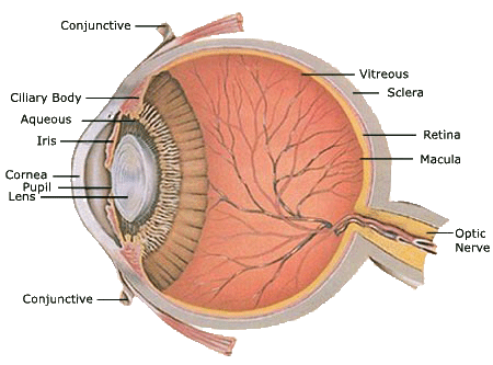
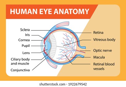
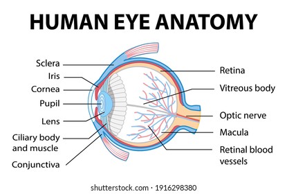
Eye Diagram With Labels and detailed description - BYJUS A brief description of the eye along with a well-labelled diagram is given below for reference. Well-Labelled Diagram of Eye The anterior chamber of the eye is the space between the cornea and the iris and is filled with a lubricating fluid, aqueous humour. The vascular layer of the eye, known as the choroid contains the connective tissue.
Human Visual Perception & Photography
Premium Vector | Diagram of human eye anatomy with label Download this Premium Vector about Diagram of human eye anatomy with label, and discover more than 37 Million Professional Graphic Resources on Freepik. #freepik #vector #eyeanatomy #cornea #retina
Eye Anatomy: imagens, fotos e vetores stock | Shutterstock
Label the Eye Worksheet - Teacher-Made Learning Resources - Twinkl In this resource, you'll find a 2-page PDF that is easy to download, print out, and use immediately with your class. The first page is a labelling exercise with two diagrams of the human eye. One is a view from the outside, and the other is a more detailed cross-section. Challenge learners to label the parts of the eye diagram. On the second page, you'll find a set of answers showing ...
picture front of the eye without labels clipart - Clipground
PDF Eye Anatomy Handout - National Institutes of Health of light entering the eye. Lens: The lens is a clear part of the eye behind the iris that helps to focus light, or an image, on the retina. Macula: The macula is the small, sensitive area of the retina that gives central vision. It is located in the center of the retina. Optic nerve: The optic nerve is the largest sensory nerve of the eye.

Anatomy - កូនខ្មែរសំរាប់អ្នក
File:Diagram of human eye without labels.svg - Wikimedia File:Diagram of human eye without labels.svg. Size of this PNG preview of this SVG file: 410 × 430 pixels. Other resolutions: 229 × 240 pixels | 458 × 480 pixels | 732 × 768 pixels | 976 × 1,024 pixels | 1,953 × 2,048 pixels.

Eye - Labelled Diagram Of Human Eye | Transparent PNG Download #836865 - Vippng
diagram of eye with labels Horseshoe Crab Anatomy. 16 Pics about Horseshoe Crab Anatomy : Label the Eye, Eye With Labels Clip Art at Clker.com - vector clip art online, royalty and also Muscles of the Human Eyeball | ClipArt ETC. Horseshoe Crab Anatomy dnr.maryland.gov crab horseshoe anatomy eyes diagram labeled gills ccs dnr maryland gov
picture front of the eye without labels clipart - Clipground
Human eye - Wikipedia Schematic diagram of the human eye. It shows a horizontal section through the right eye. The eye is made up of three coats, or layers, enclosing various anatomical structures. The outermost layer, known as the fibrous tunic, is composed of the cornea and sclera, which provide shape to the eye and support the deeper structures.

Human Body Anatomy Basics No Lines Clip Art at Clker.com - vector clip art online, royalty free ...
PDF Parts of the Eye - National Institutes of Health Eye Diagram Handout Author: National Eye Health Education Program of the National Eye Institute, National Institutes of Health Subject: Handout illustrating parts of the eye Keywords: parts of the eye, eye diagram, vitreous gel, iris, cornea, pupil, lens, optic nerve, macula, retina Created Date: 12/16/2011 12:39:09 PM
Similar Images, Stock Photos & Vectors of Human eye anatomy infographics with outside view and ...
heart diagram without labels heart labeled diagram human. Free Unlabeled Heart Diagram, Download Free Clip Art, Free Clip Art On clipart-library.com. heart diagram human simple drawing sketch blank circulatory system labels unlabeled flow easy blood worksheet label clip anatomy circulation library. 32 Label The Diagram Of The Heart - Labels Database 2020 otrasteel.blogspot.com

Diagram Of The Eye Not Labeled - Aflam-Neeeak
Label Parts of the Human Eye - University of Dayton Parts of the Eye. Select the correct label for each part of the eye. The image is taken from above the left eye. Click on the Score button to see how you did. Incorrect answers will be marked in red. ...
Human Skeleton Back No Text No Color Clip Art at Clker.com - vector clip art online, royalty ...
The Human Eye - Diagram, Parts, Working, Function and Work of The Lens The cornea, iris, pupil, and lens make up the front of the eye, which focuses the image onto the retina. The light-sensitive membrane that covers the back of the eye is known as the retina. This membrane is made up of millions of nerve cells that clump together behind the eye to form the optic nerve, a huge nerve. The Human Eye
The eye, rods and cones - Biology Notes for IGCSE 2014
Eye Diagram Teaching Resources | Teachers Pay Teachers The Human Eye Overview Reading Comprehension and Diagram Worksheet. by. Teaching to the Middle. 4.7. (65) $1.50. Zip. This passage briefly describes the human eye (900-1000 Lexile). 14 questions (matching and multiple choice) assess students' understanding. Students label a diagram of 6 parts of the eye.

1000+ images about Medical Information Eyes on Pinterest | Eye anatomy, Dry eye and Human eye
Eye Anatomy: Parts of the Eye and How We See Behind the anterior chamber is the eye's iris (the colored part of the eye) and the dark hole in the middle called the pupil. Muscles in the iris dilate (widen) or constrict (narrow) the pupil to control the amount of light reaching the back of the eye. Directly behind the pupil sits the lens. The lens focuses light toward the back of the eye.

Human Skeleton Blank Clip Art at Clker.com - vector clip art online, royalty free & public domain
The Human Eye Worksheets | Human eye diagram, Parts of the ... - Pinterest Description Use these simple eye diagrams to help students learn about the human eye. Three differentiated worksheets are included: 1. Write the words using a word bank 2. Cut and paste the words 3. Write the words without a word bank Labels include: eyebrow, eyelid, eyelashes, pupil, iris, and sclera. UPDATE: I've updated this product to ...

Anatomy, Physiology & Pathology of the Human Eye | what do you want to know is here

picture front of the eye without labels clipart 20 free Cliparts | Download images on Clipground ...
Post a Comment for "41 diagram of the human eye without labels"