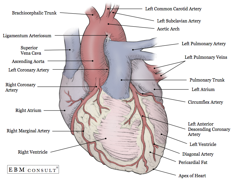
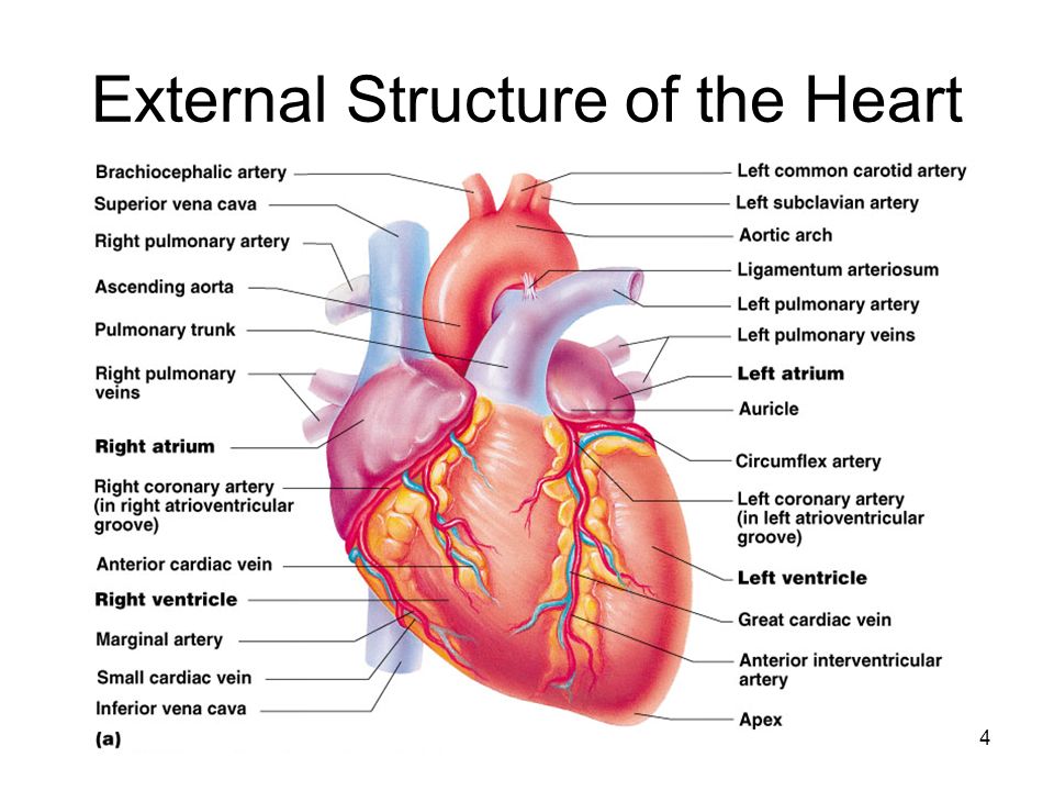
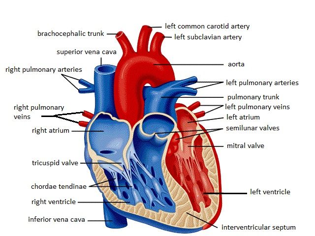
44 external structure of the heart with labels
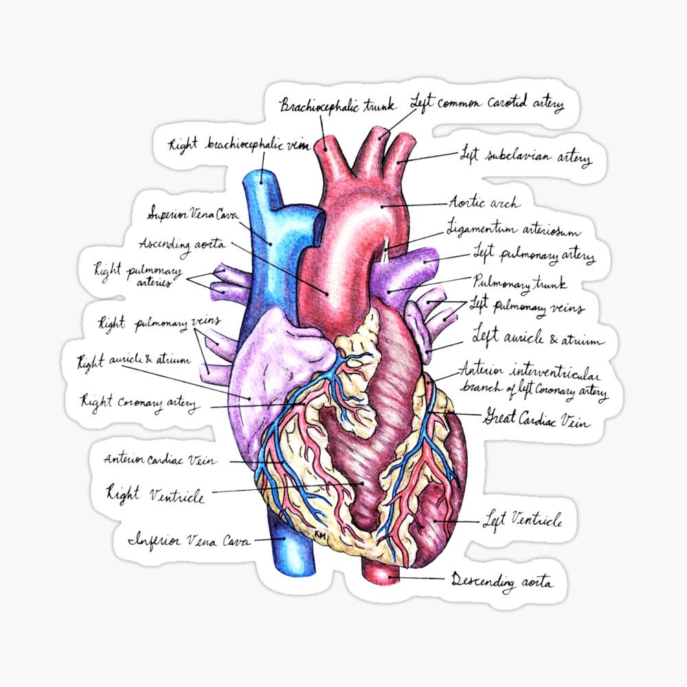
Solved External anatomy of the heart (anterior view) | Chegg.com Right coronary artery, marginal branch of RCA, Left coronary artery, circumflex branch of LCA, anterior interventricular branch of LCA, great cardiac vein Coronary blood vessels posterior view Label the following coronary arteries & veins. Also, color arteries Red, veins Blue Diagram Of Fish With Label / External Morphology Of Rohu Fish With ... The image represents the external structure of the fish and the parts are labelled. The biology of many tilapia species in natural systems is well documented. Plain diagram of the heart with labels to add and a cloze exercise on the pathway of blood through the heart. Structure of a typical fish (with diagram).
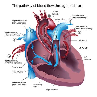
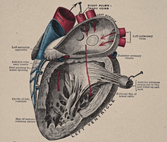
Anatomy of heart labeling Flashcards | Quizlet top of the heart right atria Receives deoxygenated blood from the body returning to the heart left atria receives oxygenated blood from the lungs via the pulmonary veins left auricle Identify the flap. right auricle Identify the flap. left ventricle Pumps oxygenated blood to the body through the aorta interatrial septum
External structure of the heart with labels
Label the heart — Science Learning Hub Label the heart Interactive Add to collection In this interactive, you can label parts of the human heart. Drag and drop the text labels onto the boxes next to the diagram. Selecting or hovering over a box will highlight each area in the diagram. pulmonary vein semilunar valve right ventricle right atrium vena cava left atrium pulmonary artery Chapter 22 Heart Flashcards | Quizlet Label the coronary arteries in an anterior view of the heart. Label the order that blood flows through in the heart, using the arrows as guides. Label the components of the heart wall. Label the components of the heart as seen from a posterior view. Label the major coronary veins. Label the components of the conduction system. External anterior heart labeling Quiz - purposegames.com This is an online quiz called External anterior heart labeling. There is a printable worksheet available for download here so you can take the quiz with pen and paper. Your Skills & Rank. Total Points. 0. ... Internal Anatomy of the Kidney 4p Image Quiz. Arteries of the abdomen view 2 6p Image Quiz. Arteries of the abdomen view 3 5p Image Quiz.
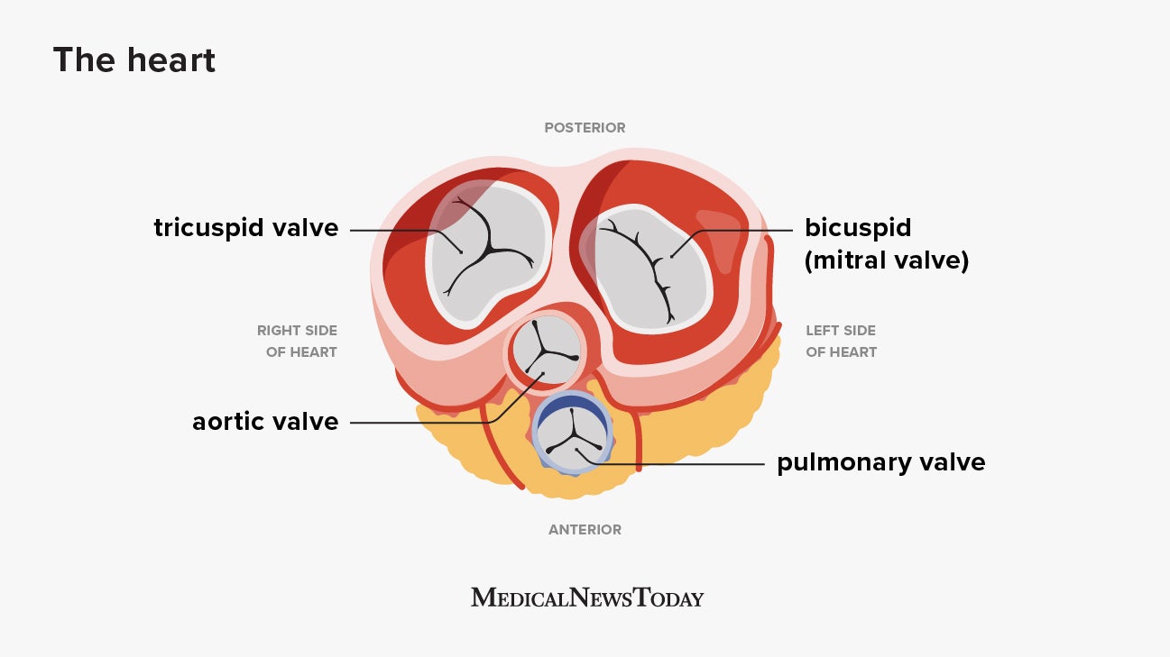
External structure of the heart with labels. EOF Cardiovascular system Diagram | Quizlet Label the coronary arteries in an anterior view of the heart. Label the internal anatomy of the heart. Match the heart valve with its description. 1. Between left ventricle and ascending aorta. 2. Between left atrium and left ventricle. 3. Between right atrium and right ventricle. Chapter 20-Cardiovascular System Flashcards | Quizlet Place the labels in order denoting the flow of oxygenated blood through the heart beginning with the vessels that bring blood back to the heart from the lungs. Correctly label the following coronary blood vessels of the heart. Correctly label these structures in this superior view of the heart. Label ECG Chapter 19: The Heart Flashcards | Quizlet Heart Location. In the thoracic cavity, between the lungs in the mediastinum. Size, Shape and Position of the Heart. •Located in thoracic cavity. -specifically in the mediastinum. •area between lungs. -superior to diaphragm. -posterior to sternum. -2/3 of heart to the left of midsagittal plane due to the liver taking space on the right.
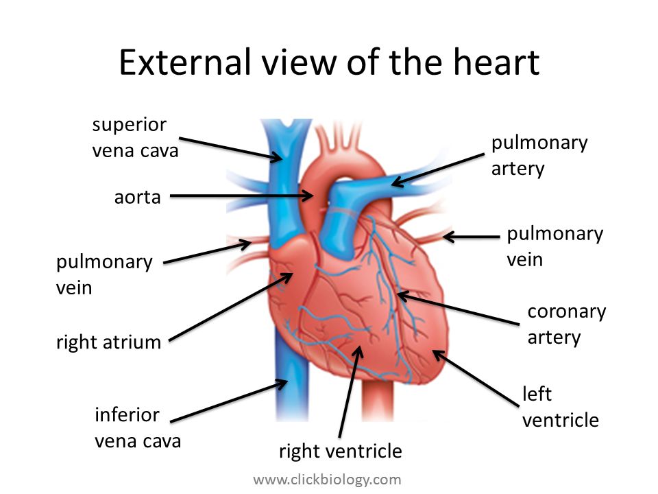
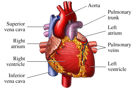
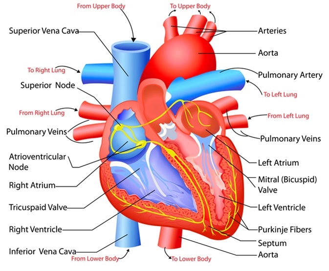
Ch. 19 Circulatory System- heart Flashcards | Quizlet Correctly label the external anatomy of the anterior heart. Place the labels in order denoting the flow of blood through the pulmonary circuit beginning with the right atrium and ending in the left atrioventricular valve. The first and last structures are given. Right atrium 1. tricuspid valve 2. right ventricle 3. pulmonary valve Lesson | The Heart - External Structure | Encounter Edu In this lesson students begin their exploration of the circulatory system, labelling a diagram of the external structures and identifying arteries and veins. They will go on to explain where blood enters and leaves the heart. Learning outcomes Structure and Function of the Heart - News-Medical.net Structure of the heart. The heart wall is composed of three layers, including the outer epicardium (thin layer), middle myocardium (thick layer), and innermost endocardium (thin layer). The ... Heart Diagram with Labels and Detailed Explanation - BYJUS Diagram of Heart. The human heart is the most crucial organ of the human body. It pumps blood from the heart to different parts of the body and back to the heart. The most common heart attack symptoms or warning signs are chest pain, breathlessness, nausea, sweating etc. The diagram of heart is beneficial for Class 10 and 12 and is frequently ...
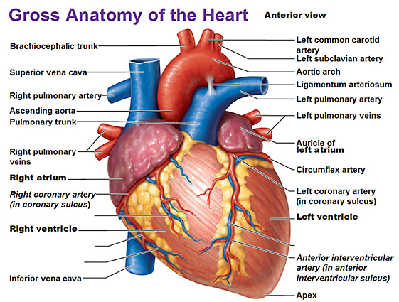
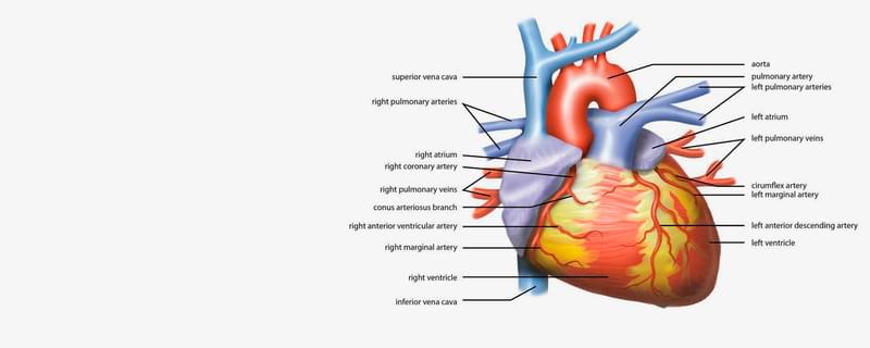
Heart anatomy: Structure, valves, coronary vessels | Kenhub The heart is shaped as a quadrangular pyramid, and orientated as if the pyramid has fallen onto one of its sides so that its base faces the posterior thoracic wall, and its apex is pointed toward the anterior thoracic wall. Structure of the Heart | SEER Training - National Cancer Institute The human heart is a four-chambered muscular organ, shaped and sized roughly like a man's closed fist with two-thirds of the mass to the left of midline. The heart is enclosed in a pericardial sac that is lined with the parietal layers of a serous membrane. The visceral layer of the serous membrane forms the epicardium. Layers of the Heart Wall Heart Anatomy: Heart Dissection - University of Washington The letters indicated in the text refer to the labels on the picture. The anterior surface of the heart is characterized by the presence of the large arteries leaving the base of the heart, the pulmonary trunk (H) and the aorta (G). The pulmonary trunk is the vessel that divides to give rise to the two pulmonary arteries going to each lung. External anterior heart labeling Quiz - purposegames.com This is an online quiz called External anterior heart labeling. There is a printable worksheet available for download here so you can take the quiz with pen and paper. Your Skills & Rank. Total Points. 0. ... Internal Anatomy of the Kidney 4p Image Quiz. Arteries of the abdomen view 2 6p Image Quiz. Arteries of the abdomen view 3 5p Image Quiz.
Anatomy of the Human Heart - Physiopedia
Chapter 22 Heart Flashcards | Quizlet Label the coronary arteries in an anterior view of the heart. Label the order that blood flows through in the heart, using the arrows as guides. Label the components of the heart wall. Label the components of the heart as seen from a posterior view. Label the major coronary veins. Label the components of the conduction system.

Important Drawings for Inter Exams | How to Draw a External Structure of The HEART | #Zoology
Label the heart — Science Learning Hub Label the heart Interactive Add to collection In this interactive, you can label parts of the human heart. Drag and drop the text labels onto the boxes next to the diagram. Selecting or hovering over a box will highlight each area in the diagram. pulmonary vein semilunar valve right ventricle right atrium vena cava left atrium pulmonary artery

मानव हृदय | external structure of heart | structure of Heart in Hindi | human heart |biology science
Heart Structure. - ppt video online download

external heart structure diagram 1 Diagram | Quizlet

heart | Structure, Function, Diagram, Anatomy, & Facts ...
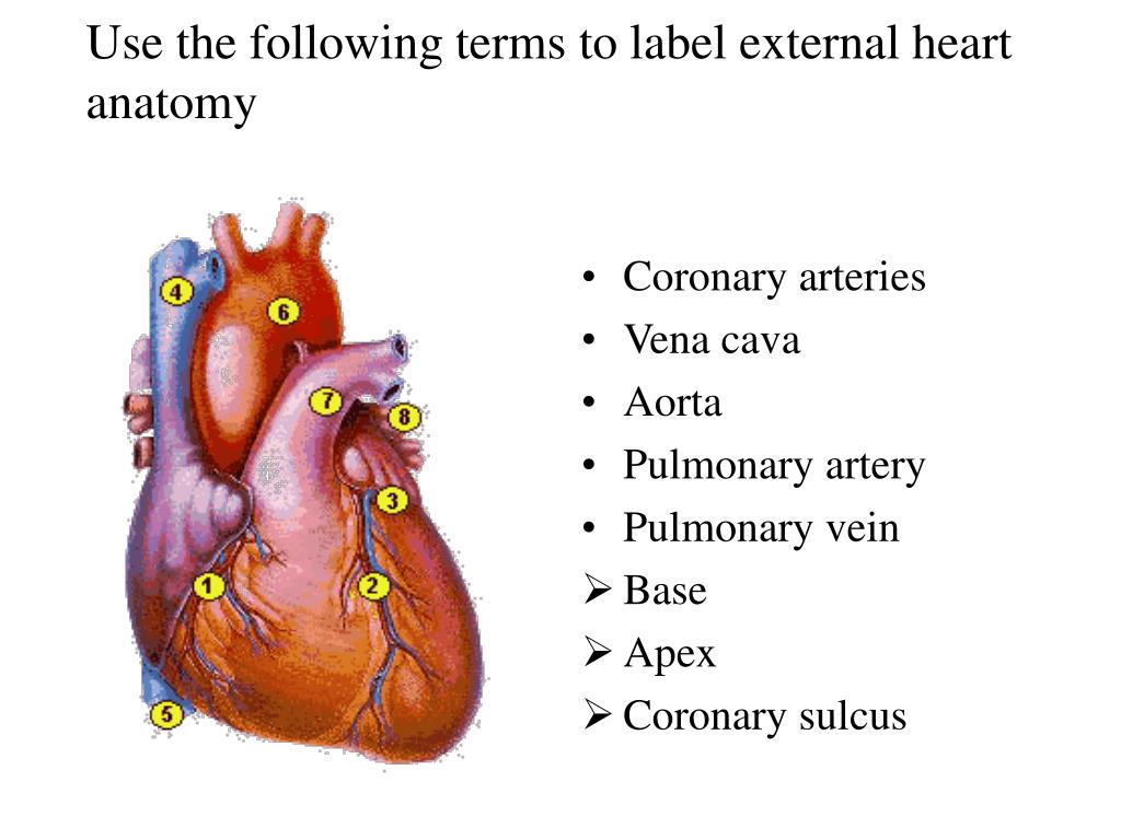
PPT - Use the following terms to label external heart anatomy ...

Form 2 Biology lesson 16 The Structure and function of the mammalian heart external structure
Label the heart — Science Learning Hub

Heart Anatomy: Labeled Diagram, Structures, Blood Flow ...
Anatomy: Heart (External)

How to Draw a External Structure of The HEART | Human body organs for kids | HumanHeartDiagram
The Cardiovascular System
Heart Anatomy: Labeled Diagram, Structures, Blood Flow ...

Heart Anatomy: Labeled Diagram, Structures, Blood Flow ...

Important Drawings for Inter Exams | How to Draw a External Structure of The HEART | #Zoology

Biology 20 Labelling the Heart
Conduction system of the heart: Parts and Functions | Kenhub
Heart - front view: MedlinePlus Medical Encyclopedia Image
Heart Anatomy
Diagrams, quizzes and worksheets of the heart | Kenhub

STRUCTURE OF HEART OF FROG || BY PHANINDRA GUPTA

What is the external structure of the human heart? - Quora
Heart: Anatomy and Function

Heart Anatomy | Anatomy and Physiology II

Heart - Wikipedia

Short / Long type answer type questions.Draw a diagram to ...

Science Is Art
SkaggsFamily.net - Dilated Cardiomyopathy

Simple heart diagram | Simple heart diagram labeled | Human ...
Heart Diagram – 15+ Free Printable Word, Excel, EPS, PSD ...
The heart: Anatomy, how it works, and more
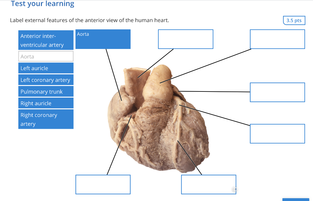
Solved Test your learning Label external features of the ...

The human heart (External and internal structure) - Online ...
:max_bytes(150000):strip_icc()/heart_interior-570555cf3df78c7d9e908901.jpg)
The 3 Layers of the Heart Wall
Structure and Function of the Heart

End of chapter exercises | Transport systems in animals ...
/the-heart-wall-4022792-FINAL-ff0aca97377c4fe9aeef72b044138011.png)
The 3 Layers of the Heart Wall
Free Unlabelled Diagram Of The Heart, Download Free ...
Lesson | The Heart - External Structure | Encounter Edu
Free Unlabelled Diagram Of The Heart, Download Free ...

File:Diagram of the human heart (cropped).svg - Wikimedia Commons

Pin on Homeschool Idea's & C.C/Ch.A Help
External Heart" Poster for Sale by KMogen5 | Redbubble
Post a Comment for "44 external structure of the heart with labels"