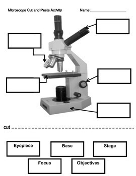
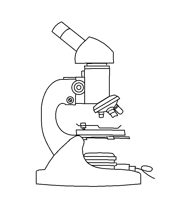
44 microscope diagram without labels
X-ray crystallography - Wikipedia X-ray crystallography is the experimental science determining the atomic and molecular structure of a crystal, in which the crystalline structure causes a beam of incident X-rays to diffract into many specific directions. By measuring the angles and intensities of these diffracted beams, a crystallographer can produce a three-dimensional picture of the density of electrons within the crystal. Icahn School of Medicine at Mount Sinai - New York City The Icahn School of Medicine. We are committed to promoting and supporting diversity and inclusion throughout our research, clinical, and educational realms among students, faculty, and staff, and in the communities we serve. The Icahn School of Medicine. The Icahn School of Medicine.
Home - Medical Marijuana, Inc. Medical marijuana refers to using the whole cannabis plant, or the plant's basic extracts, for the treatment of various ailments or conditions. If you're not treating ailments or conditions, marijuana can't be labeled medical marijuana. People often confuse the terms cannabis and marijuana. Cannabis is a category for a plant species that ...
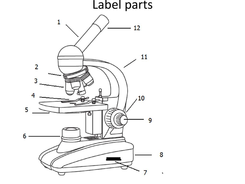
Microscope diagram without labels
Flagella: Structure, Arrangement, Function - Microbe Online Flagella (singular, flagellum) are the locomotory structures of many prokaryotes. Most protozoa and some bacteria are motile. Protozoa use flagella, cilia, or pseudopods, whereas motile bacteria move only using flagella. The flagellum functions by rotation to push or pull the cell through a liquid medium. Single-crystal X-ray Diffraction - Techniques This can be determined by viewing the samples under crossed polars on a petrographic microscope. Crystals can be broken off a larger sample and the best fragment selected. Samples should be between 30 and 300 microns, with ideal crystals averaging 150-250 microns in size. To minimize absorption affects, equant crystals are preferred. Gram Stain Technique - Amrita Vishwa Vidyapeetham Wipe the glass slide with spirit and wave the slide over the Bunsen burner to remove any unwanted microorganisms in the slide. Label one side of the glass slide with 1. Your initials 2. The date While flaming the inoculation loop be sure that each segment of metal glows orange/red-hot before you move the next segment into the flame.
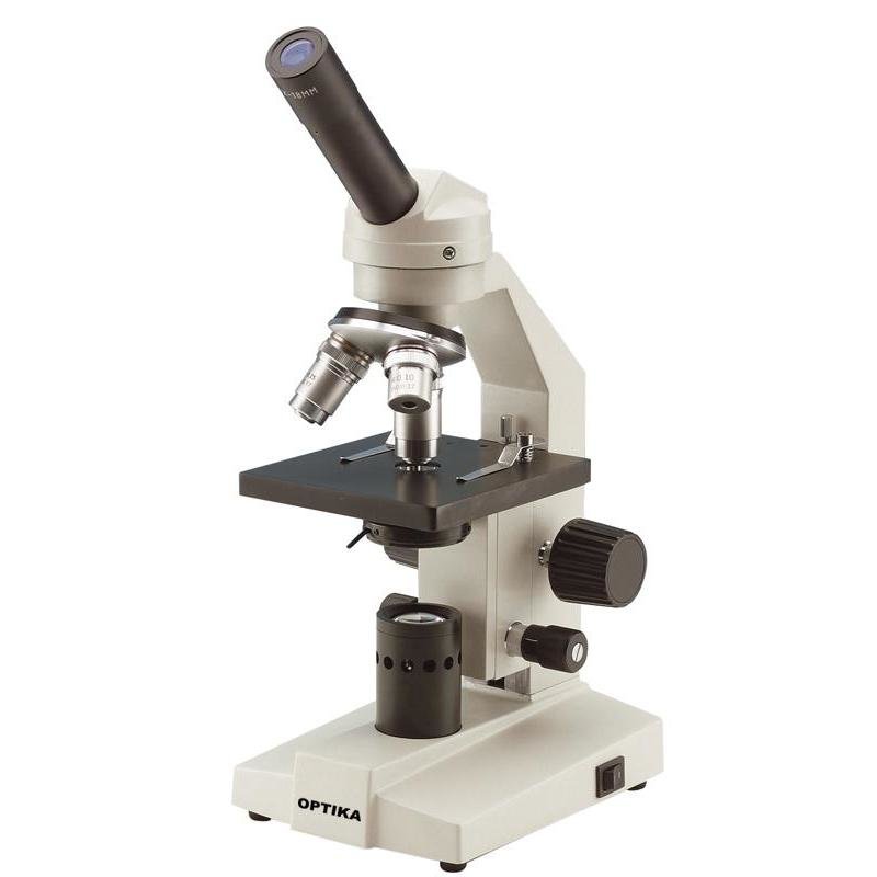
Microscope diagram without labels. achieverpapers.comAchiever Papers - We help students improve their academic ... 100% money-back guarantee. With our money back guarantee, our customers have the right to request and get a refund at any stage of their order in case something goes wrong. 11 Different Types of Microscopes (With Pictures) - Optics Mag The 11 Types of Microscopes: 1. Light Microscopes The most common type of microscope you're likely to come across, these microscopes rely on lenses and light to illuminate a specimen for optimal image-gathering. They can be used for viewing living cells, insects, for performing dissections, or for clinical blood and tissue assessment. 2. Cerebellum and brainstem: Anatomy and functions | Kenhub Anatomy of the brain (sagittal view) The cerebellum and brainstem are a testament to the fact that good things do come in small packages, so this article is an overview of their anatomy.Occupying only a fraction of the volume of the cerebrum, these structures are responsible for simplifying every second of your life and keeping you alive.Thanks to them, you can subconsciously and automatically ... Deals, Coupon Codes, Bargains & US Deals - Deals of America View More info. First login to your Amazon.com account, Now Click Here and Scroll down the landing page, at the middle of page Click on ' Clip this Coupon ' to activate $20 Off Coupon. then add ' All-Clad D3 Stainless Cookware, 12-Inch Fry Pan w/Lid ' to cart for $129.95. Your final price will be $129.95 - $20 off coupon = $109.95 + Free Shipping.
Anabaena: characteristics, habitat, reproduction and nutrition Anabaena It is a genus of prokaryotic photosynthetic cyanobacteria, that is, they are unicellular, without a defined nucleus, with genetic material, but dispersed in the cytoplasm. They are shallow water plantonic organisms, barrel-shaped and can form colonies. Cyanobacteria, including Anabaena They are also called blue-green algae, although ... Mitochondria - Genome.gov Definition. …. Mitochondria are membrane-bound cell organelles (mitochondrion, singular) that generate most of the chemical energy needed to power the cell's biochemical reactions. Chemical energy produced by the mitochondria is stored in a small molecule called adenosine triphosphate (ATP). Mitochondria contain their own small chromosomes. Bacterial Growth Curve - Amrita Vishwa Vidyapeetham Thus the increasing the turbidity of the broth medium indicates increase of the microbial cell mass (Fig 1) .The amount of transmitted light through turbid broth decreases with subsequent increase in the absorbance value. Fig 1: Absorbance reading of bacterial suspension The growth curve has four distinct phases (Fig 2) 1. Lag phase Metaphase - Genome.gov During metaphase, the nucleus dissolves and the cell's chromosomes condense and move together, aligning in the center of the dividing cell. At this stage, the chromosomes are distinguishable when viewed through a microscope. Metaphase chromosomes are used in karyotyping, a laboratory technique for identifying chromosomal abnormalities. Narration
Optical biosensing through a toy microscope over a surface 'rainbow' chip October 3, 2022 Optical biosensing through a toy microscope over a surface 'rainbow' chip by Engineering Schematic diagram of the rainbow trapping metasurface used in lung cancer diagnosis (left)... Sickle cell disease - Wikipedia Normal blood cells next to a sickle blood cell, coloured scanning electron microscope image Signs of sickle cell disease usually begin in early childhood. The severity of symptoms can vary from person to person. [15] Sickle cell disease may lead to various acute and chronic complications, several of which have a high mortality rate. [16] › articles › s41467/019/12898-9Rapid identification of pathogenic bacteria using Raman ... Oct 30, 2019 · Raman optical spectroscopy promises label-free bacterial detection, identification, and antibiotic susceptibility testing in a single step. However, achieving clinically relevant speeds and ... › scitable › topicpageFluorescence In Situ Hybridization (FISH) | Learn Science at ... (a) The basic elements of FISH are a DNA probe and a target sequence. (b) Before hybridization, the DNA probe is labeled by various means, such as nick translation, random primed labeling, and PCR.
Compound Microscope Review - ppt download
Gram Staining Procedure | New Health Advisor Label the Slides Draw a circle under the slides using a marking pen designed for glassware. This will help to designate which area to prepare the smear in the following step. You can also label them with the organism's initials at the edge of each slide. Take care that the labels do not get in contact with the reagentsused forstaining. 3.

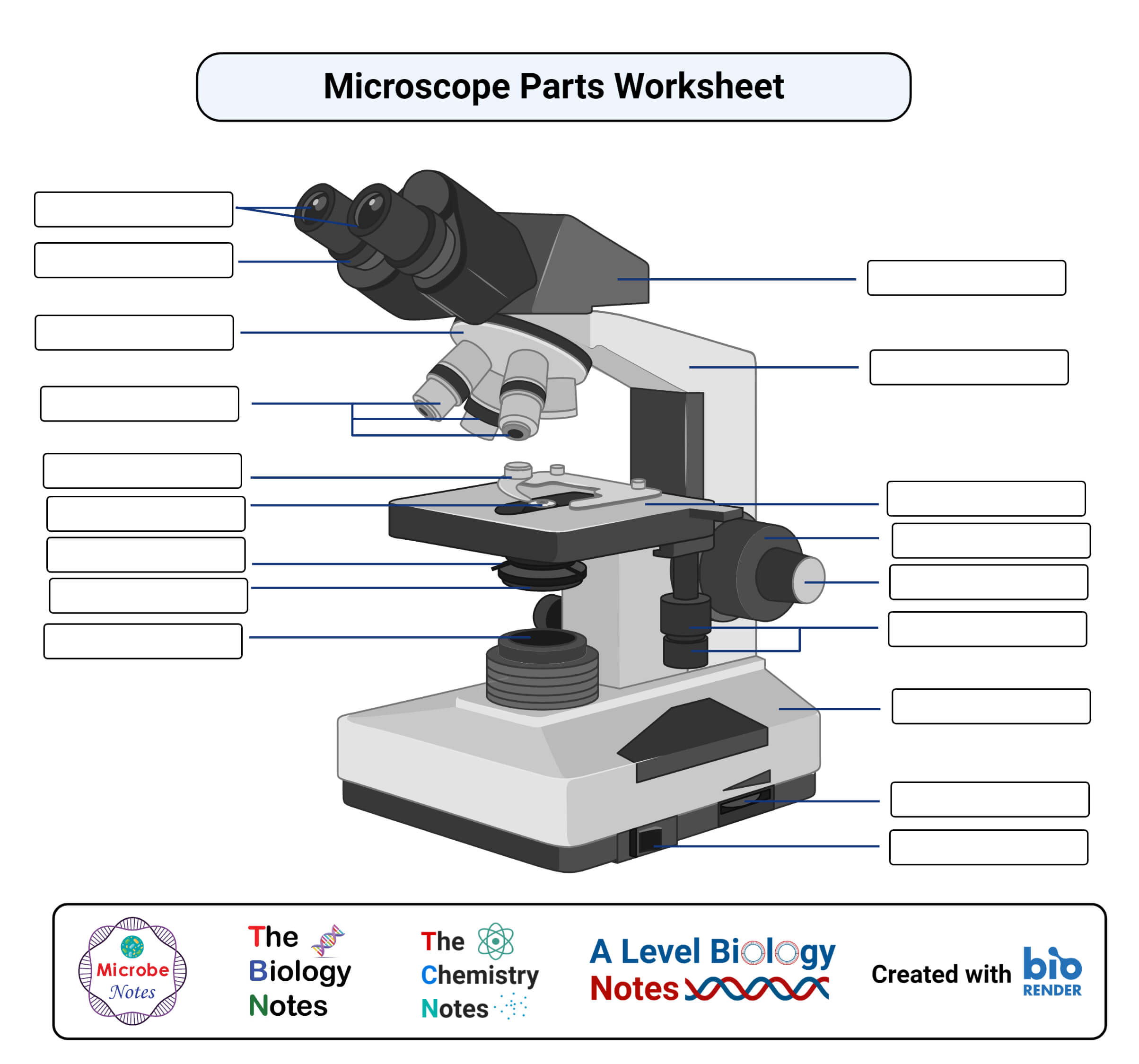
Parts of a Light Microscope Cut and Stick Worksheet - Twinkl
Emergency Showers and Eyewash Stations : OSH Answers This diameter ensures that the water will come into contact with the entire body - not just the top of the person's head. ANSI also recommends the shower head be between 208.3 and 243.8 cm (82-96 inches) from the floor. The minimum volume of spray should be 75.7 litres/minute (20 gallons/minute) for a minimum time of 15 minutes.

Simple Microscope - Diagram (Parts labelled), Principle ...
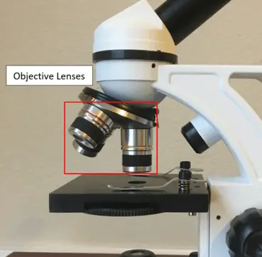
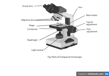
microscopeinternational.com › compound-microscopeCompound Microscope Parts, Functions, and Labeled Diagram Nov 18, 2020 · Common compound microscope parts include: Compound Microscope Definitions for Labels Eyepiece (ocular lens) with or without Pointer: The part that is looked through at the top of the compound microscope. Eyepieces typically have a magnification between 5x & 30x.

Microscope With Labels Clip Art at Clker.com - vector clip ...
Autoclave: Principle, Procedure, Types, Uses - Microbe Online After the holding period, stop the electrical heater and allow the autoclave to cool until the pressure gauge indicates that the pressure inside is equal to the atmospheric pressure. Open the discharge tap slowly and allow the air to enter the autoclave. Open the lid of the autoclave and remove the sterilized materials. Sterilization control

Parts of Stereo Microscope (Dissecting microscope) – labeled ...
Home | Daily Mail Online MailOnline - get the latest breaking news, celebrity photos, viral videos, science & tech news, and top stories from MailOnline and the Daily Mail newspaper.
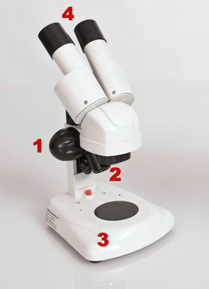
Compound and Stereo- microscopes - Microscopes 4 Schools
5 White Blood Cells Types and Their Functions - New Health Advisor 5. Basophils. Basophils are the least frequent type of white blood cell, with only 0-100 cells per mm 3 of blood. Basophils have large granules that perform functions that are not well known. They are very colorful when stained and looked at under the microscope, making them easy to identify.

Microscope labeled diagram
en.wikipedia.org › wiki › FluorescenceFluorescence - Wikipedia Fluorescence is the emission of light by a substance that has absorbed light or other electromagnetic radiation.It is a form of luminescence.In most cases, the emitted light has a longer wavelength, and therefore a lower photon energy, than the absorbed radiation.
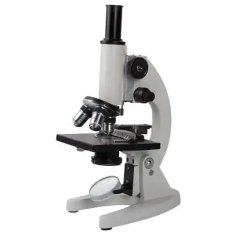
Compound Microscope - Types, Parts, Diagram, Functions and ...
Online Labs for schools - Developed by Amrita Vishwa Vidyapeetham and ... Physics, Chemistry, Biology Labs from Class 9 to Class 12. English and Maths lessons for Class 9 and 10. Interactive simulations, animations and lab videos. The concepts and understanding of the experiment. The ability to perform, record and learn experiments - anywhere, anytime, and individualised practice in all areas of experimentation.

A Study of the Microscope and its Functions With a Labeled ...
› createJoin LiveJournal Password requirements: 6 to 30 characters long; ASCII characters only (characters found on a standard US keyboard); must contain at least 4 different symbols;

Microscope Labeling Diagram | Quizlet
alex.state.al.us › plansALEX | Alabama Learning Exchange Students will use a Venn diagram to compare lightning and static electricity. Then, students will experiment with static electricity and read nonfiction passages about lightning and lightning rods. Finally, they will apply their learning to construct a model of a lightning rod system that protects a house from a lightning-induced fire.

Compound Microscope Labeled Diagram | Quizlet
Smoker's Lung Pictures: Smokers' Lungs vs. Healthy Lungs - MedicineNet Figure 1 is a diagram showing the main parts of the airway and lung. The airway consists of the oral and nasal cavities, which connect to the voice box (larynx), which connects to the windpipe (trachea). Note in the diagram that the windpipe splits into two air passages called bronchi, one going to each lung (right and left main bronchi).

label microscope diagram | Charts | Microscope, Anatomy bones ...
Mineralogy Database Complete, up-to-date, mineral database containing 4,714 mineral species descriptions and comprehensive picture library of images. These data are linked to mineral tables by crystallography, chemical composition, physical and optical properties, Dana classification, Strunz classification, mineral name origins, mineral locality information, and alphabetical listing of all known valid mineral ...
Microscope
Workplace Housekeeping - Basic Guide : OSH Answers Effective housekeeping results in: reduced handling to ease the flow of materials. fewer tripping and slipping incidents in clutter-free and spill-free work areas. decreased fire hazards. lower worker exposures to hazardous products (e.g. dusts, vapours) better control of tools and materials, including inventory and supplies.

Microscope Labeling Quiz
Gram Stain Technique - Amrita Vishwa Vidyapeetham Wipe the glass slide with spirit and wave the slide over the Bunsen burner to remove any unwanted microorganisms in the slide. Label one side of the glass slide with 1. Your initials 2. The date While flaming the inoculation loop be sure that each segment of metal glows orange/red-hot before you move the next segment into the flame.
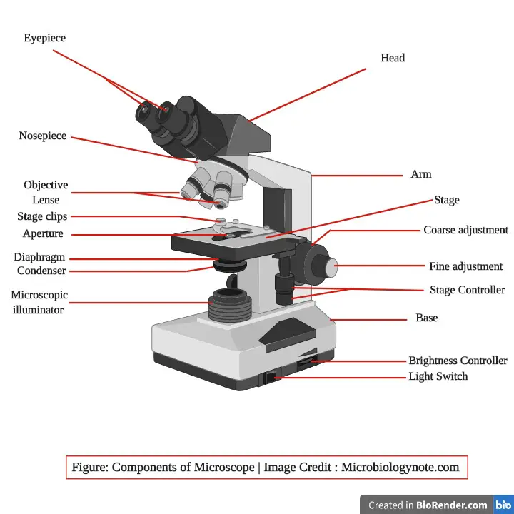
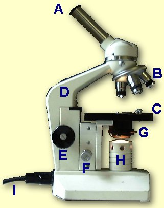
Parts of a Microscope and Their Functions
Single-crystal X-ray Diffraction - Techniques This can be determined by viewing the samples under crossed polars on a petrographic microscope. Crystals can be broken off a larger sample and the best fragment selected. Samples should be between 30 and 300 microns, with ideal crystals averaging 150-250 microns in size. To minimize absorption affects, equant crystals are preferred.

Microscope Labeling Activity - SMART Board Activity - Interactive Review
Flagella: Structure, Arrangement, Function - Microbe Online Flagella (singular, flagellum) are the locomotory structures of many prokaryotes. Most protozoa and some bacteria are motile. Protozoa use flagella, cilia, or pseudopods, whereas motile bacteria move only using flagella. The flagellum functions by rotation to push or pull the cell through a liquid medium.
How to Use the Microscope

Compound Light Microscope Labeling Diagram | Quizlet
Label a microscope - Teaching resources

Simple Microscope - Parts, Functions, Diagram and Labelling ...

The Microscope

Compound Microscope – Diagram (Parts labelled), Principle and ...

Parts of the Microscope with Labeling (also Free Printouts ...

Parts of a Compound Microscope and Their Functions

Microscope With Labels Clip Art at Clker.com - vector clip ...
The Compound Light Microscope Label the following parts on ...

Microscope - Label - Part 2 Diagram | Quizlet
Parts of a microscope with functions and labeled diagram
16 Parts of a Compound Microscope: Diagrams and Video ...
Microscope Labeling Activity

Microscope Labeling
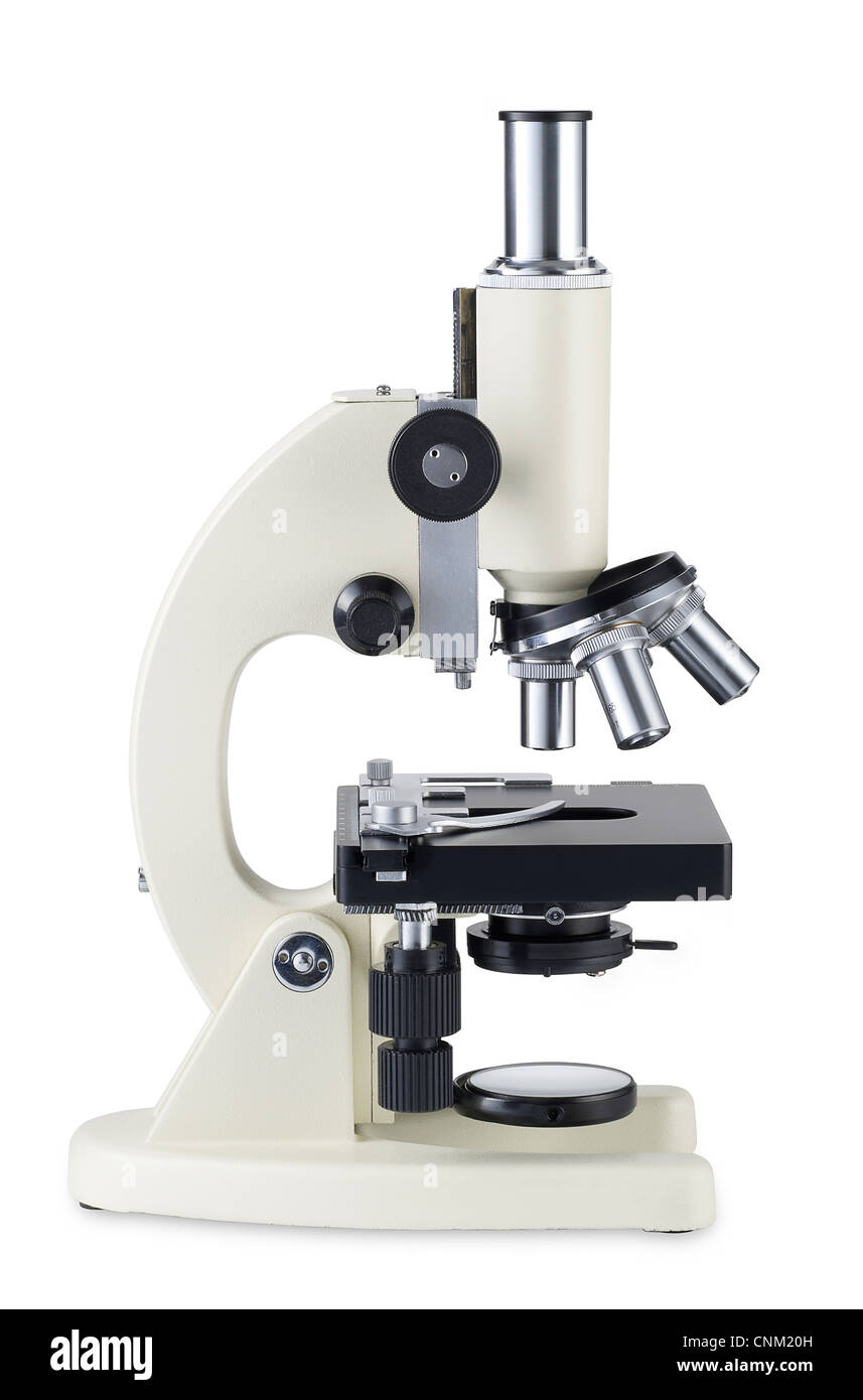
Microscope hi-res stock photography and images - Alamy
Parts of a Microscope Quiz

Label the Microscope Diagram | Download Scientific Diagram

Label the microscope — Science Learning Hub

Compound Microscope Parts, Functions, and Labeled Diagram ...
Parts of a Microscope with Their Functions – Microbe Online

Microscope World Blog: Labeling the Parts of the Microscope
unlabelled diagram of the microscope - Clip Art Library

Microscope Components - Science Quiz
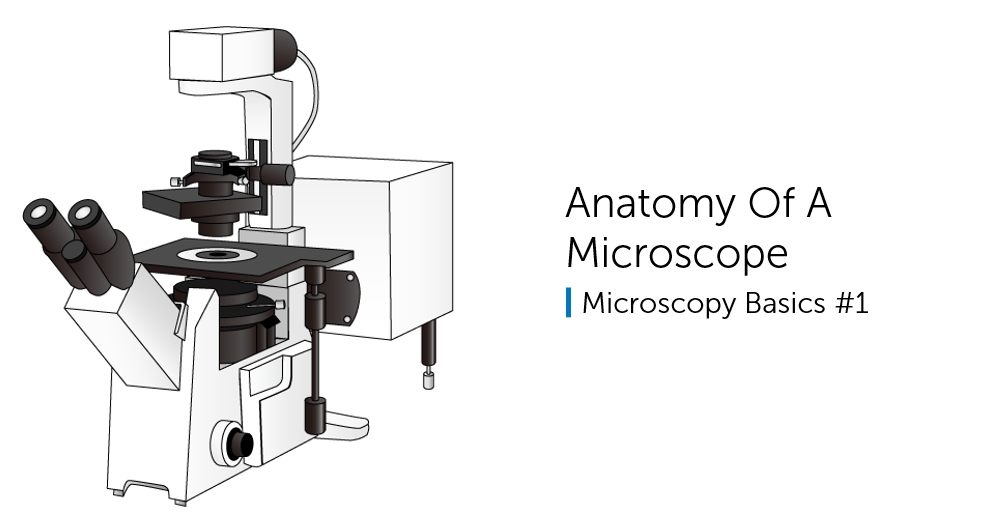
Anatomy Of A Microscope

Compound Microscope Parts – Labeled Diagram and their ...

Compound Microscope: Parts of Compound Microscope

Label a Microscope Worksheet
Post a Comment for "44 microscope diagram without labels"