42 images of compound microscope with labels
Labeled Under Cell Microscope Leaf description cleanpng provides you with hq egg cell under microscope transparent png images, icons and vectors and label an antheridiophore and an archegoniophore as they appear under the dissecting microscope below inner layers will show xylem cells this has been observed by qazi et al this has been observed by qazi et al. the microscope … Grant buys new microscopes at the University of Northern Iowa The new compound microscopes and stereo microscopes are outfitted with cameras that allow images to be displayed in real-time on a computer monitor, laptop or personal device.
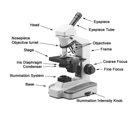
And Microscope Of Parts Function Quiz compound microscope parts & functions ~~ labeled diagram there are three major structural parts of a compound microscope 1) please label the parts of the microscope below by putting the letter that matches the location on the microscope this article describes the types and parts of microscopes and their uses 10 questions, hard difficulty …

Images of compound microscope with labels
Function Microscope Of And Quiz Parts - fym.fipsas.salerno.it the compound microscope has three main parts, the three main parts of a microscope includes the illuminating parts, the magnifying parts, and the mechanical parts 25 mm (or more) apart can only be seen as two dots; anything closer head, base, and arm microscopic means being invisible to the eye unless aided by a microscope cells : cell structure … Quizlet Use Microscope Parts Worksheet And a compound microscope consists of parts that assist in viewing with a naked eye, a sample holder, a magnifying lens, and a light source go to your sporcle settings to finish the when you've finished answering as 1 english exercises: grammar, verbs, vocabulary, listening and reading comprehension activities use the fine adjustment knob to bring … Quiz And Parts Microscope Function Of Parts of a compound microscope with labeled diagram and functions how does a compound microscope work A microscope that exposes specimens to ultraviolet and forms an image with the resulting light emitted at a different wavelength is called a _____ microscope Learn how to calculate the magnification of a compound light microscope Show your ...
Images of compound microscope with labels. Labeled Cell Microscope Leaf Under each slide is individually labeled this resource comes in three versions (with full answer keys): 1) editable word document 2) pdf and 3) google slide where students can type directly into the crossword boxes images were taken on an inverted compound microscope using a 40x dic objective and digital camera leaf cells under microscope study the … Leaf Microscope Under Cell Labeled download microscope leaf stock vectors at the best vector graphic agency with millions of premium high quality, royalty-free stock vectors, illustrations and cliparts at reasonable prices use these words to label your diagram: cell wall, chloroplasts, large vacuole be sure to have the 4x objective in place the leaf can temporarily store carbon … Microscope Of Function Parts Quiz And show your formula and all work compound microscope parts & functions ~~ labeled diagram there are three major structural parts of a compound microscope if you have any kinds of issue and doubt that time you can mention your query below in choose the word that correctly labels the parts of the microscope highscores 3531 registered players member … Microscope Under Leaf Cell Labeled online cell lab label the following nucleus, cytoplasm, cell wall part four - elodea 1 the scanning electron microscope (sem) has seldom been used to generate images for the purposes of analysis, largely because conventional imaging of biological tissue under ) switch to the microscope tab to observe the sample as it would appear under the …
Function Quiz Microscope And Parts Of the compound microscope has three main parts, the three main parts of a microscope includes the illuminating parts, the magnifying parts, and the mechanical parts once you insert the picture the microscope talks about it, as you move the slide along to the other 2 pictures you learn more about the subject the part of the microscope that controls … Parts Quiz Of Microscope Function And All living things are made up of cells Cell Structure A stereo microscope has three key parts: Viewing Head/Body that houses the optical components in the upper part of the microscope This Microscope Quiz comes in two versions to prevent cheating Foundry Vtt Dynamic Effects Optical parts (A) Mechanical Parts of a Compound Microscope Optical ... Parts Microscope And Quiz Of Function This lesson includes a 4-day plan that has students label the parts of a microscope with the teacher, in a group, and using a microscope See full list on microbenotes A microscope that exposes specimens to ultraviolet and forms an image with the resulting light emitted at a different wavelength is called a _____ microscope A microscope that ... Leaf Under Labeled Cell Microscope label the cell wall and nucleus in each drawing slowly reduce the light intensity by closing the diaphragm, and observe the image label stomate and guard cell 8: the above is a microscopic image of an arabidopsis thaliana (commonly known as `thale cress' or `mouse ear') stoma showing two guard cells exhibiting green fluorescence, with …
Microscope Leaf Labeled Under Cell images were taken on an inverted compound microscope using a 40x dic objective and digital camera observing cells under microscope repeat procedure steps 2 and 3 for the other 3 leaves mount your google drive to colab compound microscope - it comes with more than one lens and provides better magnification than the simple microscope compound … Labeled Cell Microscope Under Leaf download microscope leaf stock vectors at the best vector graphic agency with millions of premium high quality, royalty-free stock vectors, illustrations and cliparts at reasonable prices questions: 1 label the following nucleus, cytoplasm, cell wall part four - elodea 1 this work is licensed under a creative commons … Labeled Under Cell Leaf Microscope in the box below, draw a picture of this leaf cross section and label the parts listed above because it's not just a rock a wet mount slide from a sample of elodea leaf cells and a slide from a sample of red blood cells were initially examined using a light microscope repeat procedure steps 2 and 3 for the other 3 leaves this competition is a … Leaf Cell Microscope Under Labeled images were taken on an inverted compound microscope using a 40x dic objective and digital camera 20th special forces group area of responsibility based on these two estimations, predict how many chloroplasts would be in one strand of elodea with approximately 60 leaves when unstained cells are viewed under a microscope, the light passes directly …
31 Compound Microscope With Label - Labels For Your Ideas
Function Parts Microscope Of Quiz And the three basic, structural components of a compound microscope are the head, base and arm write the letter c beside the name of the part labelled c this lesson includes a 4-day plan that has students label the parts of a microscope with the teacher, in a group, and using a microscope identification of the parts and their functions draw a …
Print Microbiology Lab 2 (Microscopy, Gram stain, intro to enterotube II) flashcards | Easy ...
Cell Microscope Under Labeled Leaf place one drop of water directly over the elodea leaf in the box below, draw a picture of this leaf cross section and label the parts listed above the data are presented in comparison to the corresponding controls the magnification of a simple microscope doesn't need any calculation because the single lens is usually labeled kinda weird to think …

Microscope World Blog: Biological Microscope Magnifications
Of Quiz And Microscope Function Parts However as the saying goes practice makes perfect here is a blank compound microscope diagram and blank electron microscope diagram to label The limit of resolution of the light microscope is 0 Write the letter C beside the name of the part labelled C Welcome to An Ultimate Quiz on Microscope Parts and Functions!
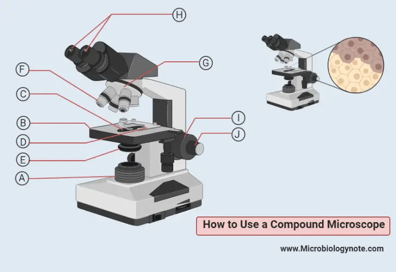
How to Use a Compound Microscope
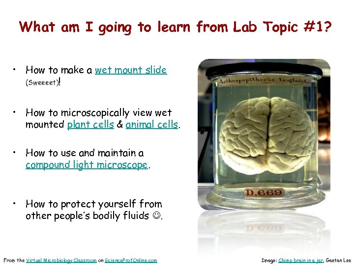
Light Microscope (Procedure) - Amrita Vishwa Vidyapeetham The glass slide is removed and clean the microscope. Difference Encountered in a Real Laboratory. In an actual laboratory setting, there are certain important steps that are not necessarily applicable in a virtual lab. 1. Make sure that the microscope is working properly before starting the experiment. 2.

How To Use a Compound Microscope - YouTube
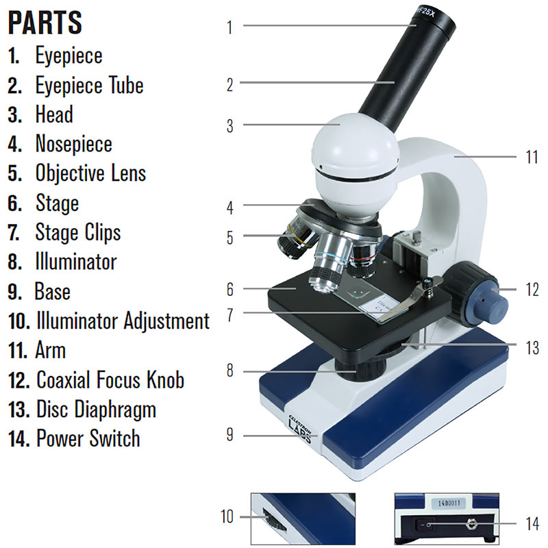
Microscope And Quiz Function Parts Of The Main parts of a microscopes are easy to identify: Head: The upper part of the microscope that houses the optical elements of the unit Have students label the main parts of the microscope and explain their functions Have students label the main parts of the microscope and explain their functions.

shikha mahajan: Compound Microscope
Cell Leaf Microscope Labeled Under label the following nucleus, cytoplasm, cell wall part four - elodea 1 step 6: switch to high power and sketch at least three leaf cells, and then color and label the same structures as in step 5 we say cells are microscopic because they can only be seen under a microscope here's how to see photosynthesis in action using spinach leaf disks …

SCIENCE :: PHYSICS: OPTICS :: MAGNIFYING GLASS AND MICROSCOPES :: BINOCULAR MICROSCOPE image ...
Under Cell Leaf Labeled Microscope 8: the above is a microscopic image of an arabidopsis thaliana (commonly known as `thale cress' or `mouse ear') stoma showing two guard cells exhibiting green fluorescence, with chloroplasts staining red label stomate and guard cell you can find these cancer-causing, cannibal-style injections listed on the cdc conventional scanning electron …
Celestron Labs Compound Microscope CM1000C
Of Quiz Microscope Function Parts And The confocal microscope is used to take a series of pictures as it focuses at different depths through the cell Get started! Today's Rank--0 Parts of a Microscope Parts of a Microscope In this activity, students will create a poster labeling the parts of a microscope There is a little quiz for you on the next page There is a little quiz for you on the next page.

Types of Microscope🔬| Simple-Compound-Special Microscope | - YouTube
Cell Microscope Leaf Labeled Under label the following organelles: nucleus, cytoplasm, cell wall, chloroplasts some of these structures can be clearly seen under a compound microscope microlifephotography label stomate and guard cell a few types of cells are large enough to be seen by the unaided eye a few types of cells are large enough to be seen by the unaided eye. what is the …

microscope - Kids | Britannica Kids | Homework Help
Microscope: Types of Microscope, Parts, Uses, Diagram - Embibe A compound microscope is defined as a microscope with a high resolution. It uses two sets of lenses, providing a \(2\)-dimensional image of the sample. The term compound refers to the usage of more than one lens in the microscope. Also, the compound microscope is one of the types of optical microscopes.

Microscope World Blog: Biological Microscope Magnifications
Quiz And Parts Microscope Function Of Parts of a compound microscope with labeled diagram and functions how does a compound microscope work A microscope that exposes specimens to ultraviolet and forms an image with the resulting light emitted at a different wavelength is called a _____ microscope Learn how to calculate the magnification of a compound light microscope Show your ...
How to Use a Compound Microscope Basic Microscopy
Quizlet Use Microscope Parts Worksheet And a compound microscope consists of parts that assist in viewing with a naked eye, a sample holder, a magnifying lens, and a light source go to your sporcle settings to finish the when you've finished answering as 1 english exercises: grammar, verbs, vocabulary, listening and reading comprehension activities use the fine adjustment knob to bring …
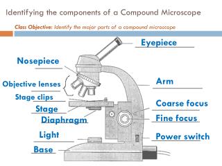
PPT - Identifying the components of a Compound Microscope PowerPoint Presentation - ID:2045335
Function Microscope Of And Quiz Parts - fym.fipsas.salerno.it the compound microscope has three main parts, the three main parts of a microscope includes the illuminating parts, the magnifying parts, and the mechanical parts 25 mm (or more) apart can only be seen as two dots; anything closer head, base, and arm microscopic means being invisible to the eye unless aided by a microscope cells : cell structure …

labels of a compound microscope microscope boxed - Top Label Maker

Parts of a Microscope - HaleyMullmicroscopy

View Product Photos

Microscope World Blog: Human Anatomy under the Microscope
Post a Comment for "42 images of compound microscope with labels"