38 diagram of the lungs with labels
Lungs (Human Anatomy): Picture, Function, Definition, Conditions - WebMD The lungs are a pair of spongy, air-filled organs located on either side of the chest (thorax). The trachea (windpipe) conducts inhaled air into the lungs through its tubular branches, called... lungs diagram to label Medical pictures info - bronchial tree. The lungs are a pair of organs within the rib cage that a.... Human lung anatomy diagram royalty free vector image , #affiliate, #. lungs diagram to label. draw a well labelled diagram of the section of an alveolus and the. 11 Images about draw a well labelled diagram of the section of an alveolus and ...
labeled diagram of the lungs labeled diagram of the lungs Respiratory System Worksheet - WikiEducator. 9 Images about Respiratory System Worksheet - WikiEducator : Respiratory System Diagram to Label Best Of Respiratory System Diagram, Respiratory System Week #8 Flashcards | Easy Notecards and also 5. Frog Dissection - Ms. Spira SNC 2P Grade 10 Applied Science.

Diagram of the lungs with labels
Drawing of lungs with labels - Jcap View Original Image at Full Size, Encourage your students to work independently and label the parts of the body they can see, mouth, Lung diagram in this post.Lung anatomy diagram or Simple lungs diagram with label are also mentioned below.We are providing simple lungs diagram for quick drawing the diagram. Labeled Diagram of the Human Lungs diagram of lungs to label lung hilum lungs anatomy medial surface diagram kenhub arteries drawing segments bronchopulmonary innervation blood Alveoli evolvingsciences.com label diagram alveoli draw membrane diffusion evolvingsciences Posterior View Angled To The Right Hand Side Of The Lungs And Ribcage Diagram Lungs Stock Illustrations - 2,588 Diagram Lungs ... - Dreamstime Download 2,588 Diagram Lungs Stock Illustrations, Vectors & Clipart for FREE or amazingly low rates! New users enjoy 60% OFF. 189,316,523 stock photos online. ... Labeled diagram with brain sections. Cranial nerves vector illustration. Labeled diagram with brain sections and its.
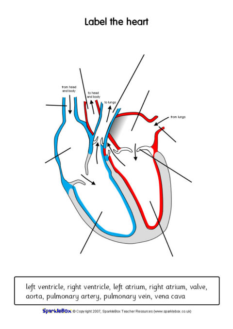
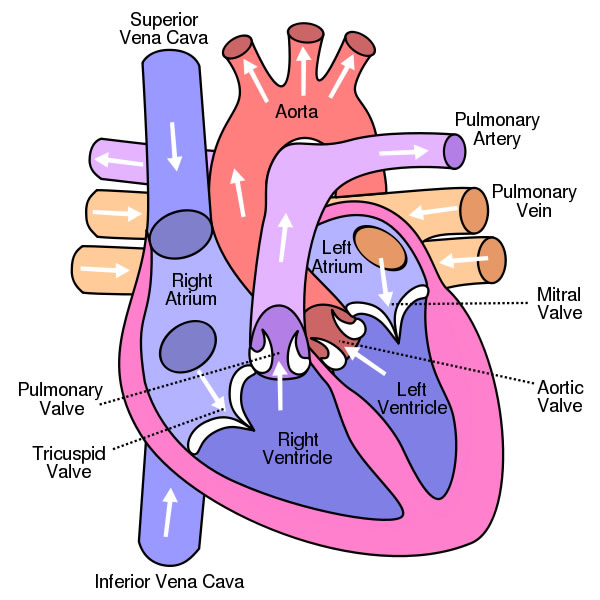
Diagram of the lungs with labels. Label the heart — Science Learning Hub Jun 16, 2017 · Drag and drop the text labels onto the boxes next to the diagram. Selecting or hovering over a box will highlight each area in the diagram. In this interactive, you can label parts of the human heart. ... Receives oxygenated blood from the lungs. Left ventricle. Region of the heart that pumps oxygenated blood to the body. Pulmonary artery ... Diagram Of The Lungs With Labels Labeling Of The Lungs Label The Lungs ... Diagram Of The Lungs With Labels Labeling Of The Lungs Label The Lungs Diagram Diagram Of Lungs With. By admin Apr 15, 2019. Share this page . Post navigation. Lung Lobectomy: What you need to know . By admin. Related Post. Leave a Reply Cancel reply. You must be logged in to post a comment. Lung Diagram Labeled | EdrawMax Template In the following lung labeled diagram, we have shown Thyroid cartilage, Cricoid Cartilage, Tracheal Cartilage, Apex, Left Upper Lobe, Hilum, Left Bronchus, Oblique Fissure, Bronchioles, Left Lower lobe, Base of lung, cardiac notch, right lower lobe, oblique fissure, right middle lobe, horizontal fissure, right bronchus, right upper lobe, and tra... Circulatory System Diagram - Cardiovascular System and Blood ... They may come with or without labels. Common circulatory system diagrams show pulmonary circulation, coronary circulation, systematic circulation, veins, arteries, or a combination. The systemic circulation system is the most commonly illustrated of the systems that make up the circulatory system as it is the largest.
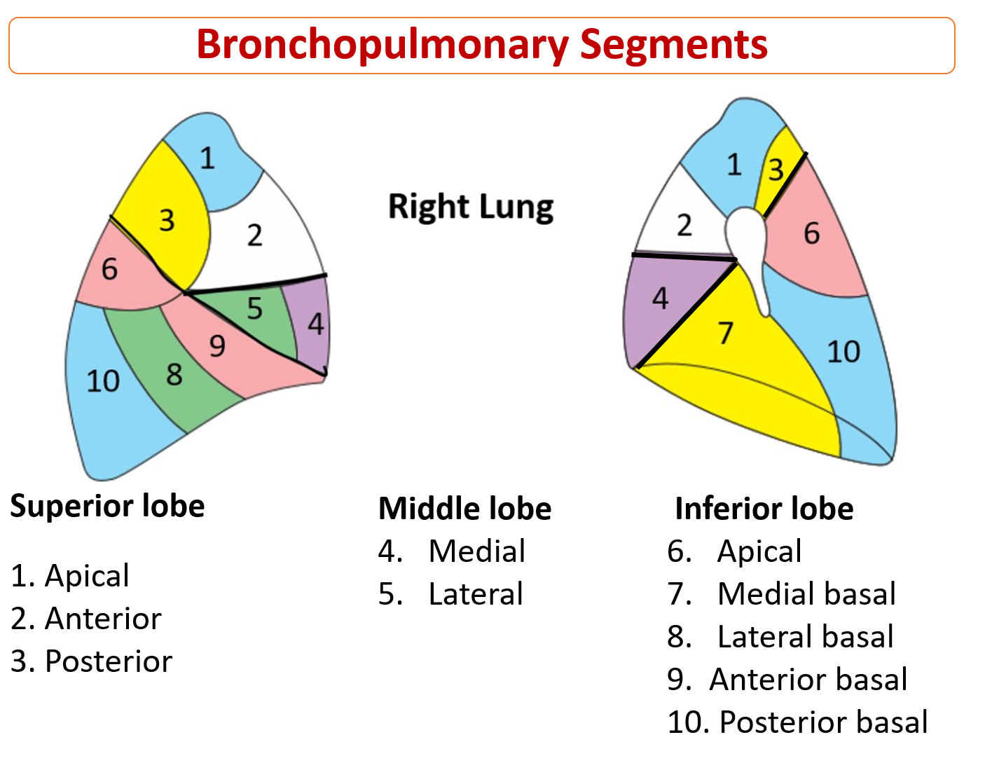
Lung Diagram | Free Lung Diagram Template - Edrawsoft The lung diagram template here clearly presents a pair of spongy on both side of the chest. Simply hitting on the template to learn more parts including pleura, ribs, bronchi, alveoli and more. Feel free to find out more human anatomy templates and symbols in the free download version. A Guide to Understand Lung with Diagrams | EdrawMax Online Lobes (two or three): The fissures on the lungs separate them into lobes. The right lung has three lobes (superior, middle, and inferior), while the left has two (superior and inferior). The lobes of the right lung are divided by fissures, namely oblique and horizontal fissures. An oblique fissure separates the lobes of the left lung. human respiratory system diagram labeled diagram system labels heart respiratory worksheet anatomy animals blank trachea structures alveoli physiology diaphragm pleural ribs cavity list cartilage rings. Structure Of The Lungs | ClipArt ETC etc.usf.edu. lungs clipart system human respiratory structure lapbook body etc medium diagrams. Thymus Gland Of Endocrine System. Fully Labelled Diagram Alveolus Lungs Showing Stock ... - Shutterstock Shutterstock customers love this asset! Stock Vector ID: 369984683 Fully labelled diagram of the alveolus in the lungs showing gaseous exchange. Vector Formats EPS 1114 × 800 pixels • 3.7 × 2.7 in • DPI 300 • JPG Show more Vector Contributor S Steve Cymro Similar images See all Assets from the same collection Similar video clips
Labeled Diagram of the Human Lungs - Bodytomy Given below is a labeled diagram of the human lungs followed by a brief account of the different parts of the lungs and their functions. Each lung is enclosed inside a sac called pleura, which is a double-membrane structure formed by a smooth membrane called serous membrane. Female Anatomy Stock Photos, Pictures & Royalty-Free Images Female reproductive system with image diagram Female reproductive system with image diagram female anatomy stock pictures, royalty-free photos & images ... Human anatomy scientific illustrations with latin/italian labels: female reproductive organ. ... Woman silhouette with lungs, heart, thyroid, stomach, liver, kidneys, uterus, intestine ... DIAGRAMS: Lungs Cross Section - Labeled - Body Diagram | Body diagram ... Human Anatomy. Shortness of breath when not performing a strenuous activity is a symptom every woman should take seriously. Difficulty breathing can be an indicator of several disease processes, ranging from: infections like pneumonia and bronchitis pulmonary problems such as asthma blood clots in the lungIt also... 3. 30Seconds. drawing of lungs with labels File:Diagram of the human heart (cropped).svg - Wikimedia Commons we have 9 Pictures about File:Diagram of the human heart (cropped).svg - Wikimedia Commons like Diagram of Air Tubes in the Lungs | ClipArt ETC, Respiratory System With Label Drawing at GetDrawings.com | Free for and also Simple Pavement Epithelium Cells | ClipArt ETC.

Cat Dissection
Heart Diagram with Labels and Detailed Explanation - BYJUS The diagram of heart is beneficial for Class 10 and 12 and is frequently asked in the examinations. A detailed explanation of the heart along with a well-labelled diagram is given for reference. ... The pulmonary artery, being an exception, carries deoxygenated blood to the lungs for purification. The veins carry impure blood from different ...
Diagram of Circulation | ClipArt ETC
How to draw and label a lung | step by step tutorial - YouTube A beautiful drawing of Lung. And it will teach you to draw the lung very easily. Watch the video and please be kind enough to thumbs up my videos. And I will...

Lifewords
Human Throat Anatomy Pictures, Images and Stock Photos Human Respiratory System anatomical vector illustration, medical education cross section diagram with nasal cavity, throat, lungs and alveoli. Human Respiratory System anatomical vector illustration, medical education cross section diagram with nasal cavity, throat, esophagus, trachea, lungs and alveoli. human throat anatomy stock illustrations
Lungs - Anatomy QA
Lung Anatomy, Function, and Diagrams - Healthline The lungs begin at the bottom of your trachea (windpipe). The trachea is a tube that carries the air in and out of your lungs. Each lung has a tube called a bronchus that connects to the trachea....
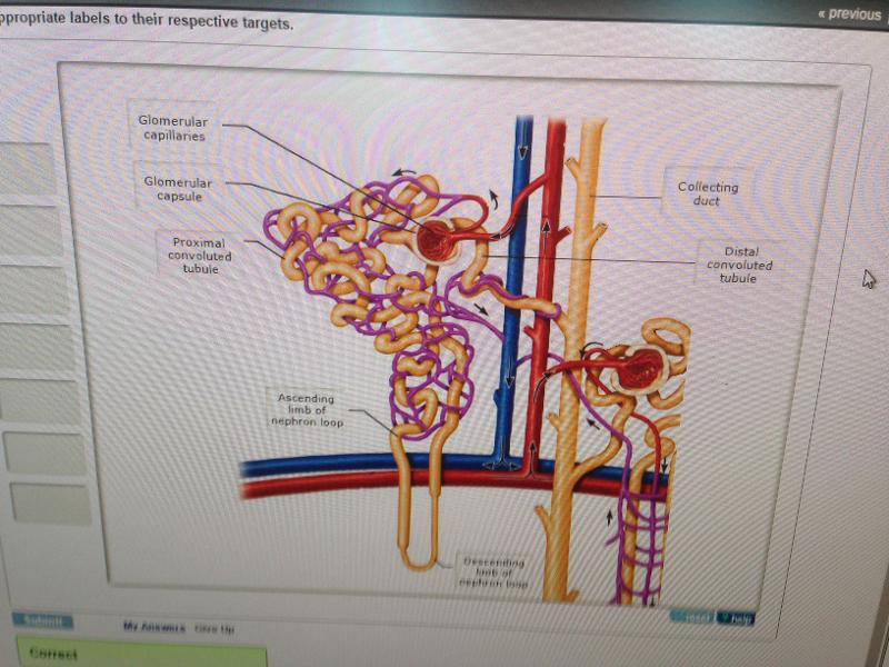
Chapter 25: The Urinary System (Mastering) Flashcards | Easy Notecards
the lungs labeled diagram the lungs labeled diagram Unlabelled Respiratory System Clip Art at Clker.com - vector clip art. 11 Images about Unlabelled Respiratory System Clip Art at Clker.com - vector clip art : Lungs Labeled Diagram Health Stock Vector - Illustration of disease, FIGURE 14.2.
Related Items
8,700 Lung diagram Images, Stock Photos & Vectors - Shutterstock 8,755 lung diagram stock photos, vectors, and illustrations are available royalty-free. See lung diagram stock video clips Image type Orientation Color People Artists Sort by Popular Healthcare and Medical Anatomy Icons and Graphics Recreation/Fitness lung respiratory system medicine pulmonary alveolus organ human body Next of 88
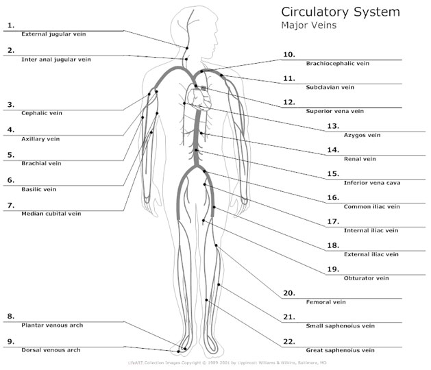
Circulatory System Diagram - Cardiovascular System and Blood Circulation Diagram
Diagram of Human Heart and Blood Circulation in It Four Chambers of the Heart and Blood Circulation. The shape of the human heart is like an upside-down pear, weighing between 7-15 ounces, and is little larger than the size of the fist. It is located between the lungs, in the middle of the chest, behind and slightly to the left of the breast bone. The heart, one of the most significant organs ...
Human Heart Diagram - Human Body Pictures - Science for Kids
Frog Dissection Guide & High School Science Lesson | HST Internally, the tadpole’s gills are replaced with lungs until finally, the tadpole has become a frog. The young frog grows and matures to adulthood over a period of 2-4 years. The adult frogs then lay their eggs and begin the cycle again. ... When you’re done, print out this diagram and fill in the labels yourself to test your knowledge of ...
Post a Comment for "38 diagram of the lungs with labels"